Anatomy/Sense Organs
Sense Organs is a topic for the event Anatomy and Physiology. It rotates in for the first time for 2017 season.
Sense Organs
Some of this information can be found on the relevant section on the Nervous System page.
Sense Organs consist of the eye, ear, nose, and tongue.
Eye
Vision is the dominant sense, with 70% of all sensory receptors in the body being in the eyes. In adults, the eye has a diameter of about one inch.
Accessory Structures
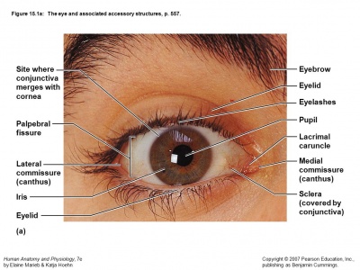
Accessory Structures of the eye include the eyebrows, eyelids, conjunctiva, lacrimal apparatus and extrinsic eye muscles.
Eyebrows
Eyebrows, the strips of hairs above the eye socket, prevent perspiration from getting into the eyes and shade the eyes from sunlight. They are short coarse hairs that overlie the supraorbital margins of the skull. They help shade the eyes from sunlight and prevent perspiration trickling down the forehead from reaching the eyes.
Eyelids
Eyelids, also known as palpebrae, provides protection to the anterior part of the eye. The superior and inferior eyelids meet at the medial and lateral canthi. Next to the medial canthus is a fleshy elevation called the lacrimal caruncle, which contains sebaceous and sweat glands. These glands produce a whitish oily secretion, most commonly known as “eye boogers”. Eyelids are supported internally by connective tissue sheets called tarsal plates.The levator palpebrae superioris muscle raises the superior eyelid and thus opens the eye, whereas the orbicularis oculi muscle brings the eyelids together and thus shuts the eye. These muscles are activated to cause blinking every 3-7 seconds to prevent dehydration of the eye. Every time we blink, accessory structure secretions are spread across the eye. These muscles are also activated when the eye is threatened by foreign objects.
Projecting from the edge of each eyelid are the eyelashes. Since they are richly innervated, anything that touches the eye triggers blinking.
Tarsal glands, also known as Meibomian glands, are located in the tarsal plates. Their ducts open just behind the eyelashes, and they produce a lubricating oily secretion called meibum. The conjunctiva is a transparent mucous membrane that lines the anterior surface of the eye, as well as the insides of the eyelid.
The lacrimal apparatus consists of the lacrimal gland and a series of ducts that drain excess lacrimal secretions (tears) into the nasal cavity. The lacrimal gland produces tears that travel through excretory ducts and onto the surface of the eye. Blinking spreads the tears to the medial commissure, where they enter the lacrimal canaliculi via two small holes called lacrimal puncta. The lacrimal canaliculi leads to the lacrimal sac and then the nasolacrimal duct. From there, the tears empty into the nasal cavity. Tears not only lubricate and moistens the eye, but it also cleanses the surface of the eye, since it contains antibodies and lysozyme, an enzyme that kills bacteria.
Conjunctiva
Is a transparent membrane that lines the eyelids and folds back over the anterior surface of the eyeball. Major function of the conjunctiva is to produce a lubricating mucus that prevents the eyes from drying out
Lacrimal Apparatus
Consist of the lacrimal gland and the ducts that drain lacrimal secretions into the nasal cavity. The lacrimal gland lies in the orbit of the eye and continually secretes a dilute saline solution called lacrimal secretion – or more commonly called tears. Lacrimal fluid contains mucus, antibodies and lysozyme (enzyme that destroys bacteria).
Extrinsic Eye Muscles
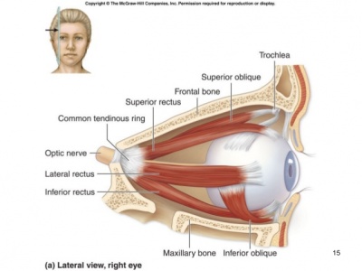
These muscles originate from the walls of the orbit and insert into the outer surface of the eyeball. They allow the eyes to follow a moving object, help maintain the shape of the eye and hold it in the orbit. Are among the most precisely and rapidly controlled skeletal muscles in the entire body.
The extrinsic eye muscles include four rectus muscles: superior, inferior, lateral, and medial, that originate from the common tendinous ring and run straight to their insertion, and two oblique muscles: superior and inferior. The superior oblique muscle also originates from the common tendinous, but runs along the medial wall of the orbit, makes a right angle turn through a loop called the trochlea, and inserts on the superior aspect of the eyeball. The inferior oblique originates from the medial orbit surface and runs laterally to insert on the the inferior aspect of the eye.
| Muscle | Action | Controlling cranial nerve |
|---|---|---|
| Lateral Rectus | Moves eye laterally | Abducens (VI) |
| Medial Rectus | Moves eye medially | Oculomotor (III) |
| Superior Rectus | Rotates eye medially and elevates eye | Oculomotor (III) |
| Inferior Rectus | Rotates eye medially and depresses eye | Oculomotor (III) |
| Inferior Oblique | Rotates eye laterally and elevates eye | Oculomotor (III) |
| Superior Oblique | Rotates eye laterally and depresses eye | Trochlear (IV) |
Structure Of The Eyeball
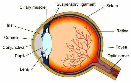
Structure of the Eyeball Its wall is composed of three layers: the fibrous, vascular and inner layers.
Fibrous Layer – the outermost coat of the eyeball and composed of dense avascular connective tissue. Vascular layer – forms the middle coat of the eyeball. Also called the uvea and contains three major regions:
Inner Layer (Retina) - the retina which develops from an extension of the brain, contains (1) millions of photoreceptors that transduce (convert) light energy, (2) other neurons involved in the processing response to light and (3) glia. Posses two layers the pigmented layer and the neural layer. Only the neural plays a part in vision.
Eye- the organ of vision. It consists of a transparent lens that focuses light on the retina. The retina is covered with light-sensitive cells-rods and cones. The cone cells are sensitive to color and are located in the part of the retina called the fovea, where the light is focused by the lens. The rod cells are not sensitive to color, but have greater sensitivity to light than the cone cells. These cells are located around the fovea and are responsible for peripheral vision and night vision. The eye is connected to the brain through the optic nerve. The optic nerve is called the "blind spot" because it is insensitive to light. The occipital lobe of the brain maps the visual input from the eyes. The brain combines the input of our two eyes into a three-dimensional image. In addition, even though the image on the retina is upside-down because of the focusing action of the lens, the brain compensates and provides the right-side-up perception. In the dark, a substance produced by the rod cells increases the sensitivity of the eye so that it is possible to detect very dim light. In strong light, the iris contracts reducing the size of the aperture that admits light into the eye and a protective obscure substance reduces the exposure of the light-sensitive cells. The spectrum of light to which the eye is sensitive varies from the red to the violet. Lower electromagnetic frequencies in the infrared are sensed as heat, but cannot be seen. Higher frequencies in the ultraviolet and beyond cannot be seen either, but can be sensed as tingling of the skin or eyes depending on the frequency. The human eye is not sensitive to the polarization of light, i.e., light that oscillates on a specific plane.
Ear
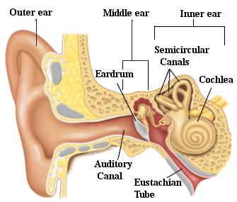
Ear- the organ of hearing. The outer ear protrudes away from the head and is shaped like a cup to direct sounds toward the tympanic membrane, which transmits vibrations to the inner ear through a series of small bones in the middle ear called the malleus, incus and stapes. The inner ear, or cochlea, is a spiral-shaped chamber covered internally by nerve fibers that react to the vibrations and transmit impulses to the brain via the auditory nerve. The brain combines the input of our two ears to determine the direction and distance of sounds. The inner ear has a vestibular system formed by three semicircular canals that are approximately at right angles to each other and which are responsible for the sense of balance and spatial orientation. The inner ear has chambers filled with a viscous fluid and small particles (otoliths) containing calcium carbonate. The movement of these particles over small hair cells in the inner ear sends signals to the brain that are interpreted as motion and acceleration. The human ear can perceive frequencies from 16 cycles per second, which is a very deep bass, to 28,000 cycles per second, which is a very high pitch. The human ear can detect pitch changes as small as 3 hundredths of one percent of the original frequency in some frequency ranges. Some people have "perfect pitch", which is the ability to map a tone precisely on the musical scale without reference to an external standard.
Nose
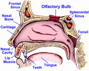
Nose- the organ of smell. The nose is the organ responsible for the sense of smell. The cavity of the nose is lined with mucous membranes that have smell receptors connected to the olfactory nerve. The smells themselves consist of vapors of various substances. The smell receptors interact with the molecules of these vapors and transmit the sensations to the brain. The nose also has a structure called the vomeronasal organ whose function has not been determined, but which is suspected of being sensitive to pheromones that influence the reproductive cycle. The smell receptors are sensitive to seven types of sensations that can be characterized as camphor, musk, flower, mint, ether, acrid, or putrid. The sense of smell is sometimes temporarily lost when a person has a cold.
Tongue
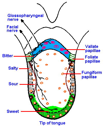
Tongue- the organ of taste. The receptors for taste, called taste buds, are situated chiefly in the tongue, but they are also located in the roof of the mouth and near the pharynx. They are able to detect four basic tastes: salty, sweet, bitter, and sour. The tongue also can detect a sensation called "umami" from taste receptors sensitive to amino acids. Generally, the taste buds close to the tip of the tongue are sensitive to sweet tastes, whereas those in the back of the tongue are sensitive to bitter tastes. The taste buds on top and on the side of the tongue are sensitive to salty and sour tastes. At the base of each taste bud there is a nerve that sends the sensations to the brain. The sense of taste functions in coordination with the sense of smell. The number of taste buds varies substantially from individual to individual, but greater numbers increase sensitivity. The gouge is covered with papillae which makes the tongue have a rough texture.


