Anatomy/Nervous System
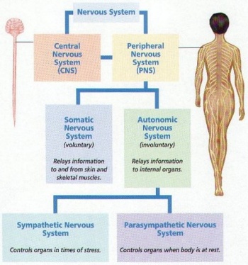
The nervous system is a focus topic of the event Anatomy and Physiology. It is expected to run in the 2022 season, and was previously a topic in 2017, 2014 and 2013. The nervous system consists of your brain and all the nerves throughout your body. It is responsible for regulating the body's response to external and internal stimuli. It can be divided into the central and peripheral nervous systems. The central nervous system or CNS includes the brain, cranial nerves, and the spinal cord; the peripheral nervous system or PNS, includes all nerves outside of the spinal region.
Parts of the Nervous System
As stated above, the nervous system consists of the central nervous system (CNS) and the peripheral nervous system (PNS).
Central Nervous System
Consists of the Brain and Spinal Cord.
- Parts of the Brain- view below
- Organization of the Spinal Cord- below
Peripheral Nervous System
Nerves and ganglia not including the CNS.
Split into afferent and efferent nervous systems:
- afferent - sensory
- efferent - motor.
The efferent nervous system is split into the autonomic and somatic nervous systems.
- autonomic - controlled by the subconscious. Happens "auto"matically. Can control all types of muscle
- somatic - controlled by the conscious brain. Always effects skeletal muscle.
The autonomic nervous system is split into the sympathetic and parasympathetic nervous systems.
- sympathetic - "fight or flight" response. Responds in times of stress.
- parasympathetic - controls the body in times of rest.
Brain and Sense Organs
There are a wide variety and a vast number of parts in the Nervous System, but the major regions are the brain, nerves, and spinal cord, all of which contribute to the great capabilities of the human body.
The Brain
Your brain is arguably the most important organ in your body. It controls all actions, stores memory, and processes the 5 senses. It has many important parts, each with a unique and vital role.
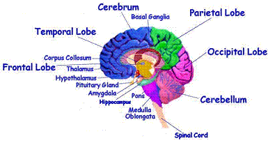
The brain is divided into four main lobes: the frontal lobe, the parietal lobe, the occipital lobe, and the temporal lobe. Each has a specific function.
Frontal Lobe
The frontal lobe is responsible for conscious thought; damage to it can result in severe personality changes. It is positioned anterior to the central sulcus. It is generally responsible for recognizing future consequences resulting from current actions, choosing between good and bad actions (or better and best), overriding and suppressing socially unacceptable responses, and determining similarities and differences between things or events. In the posterior portion of the frontal lobe lies the precentral gyrus which is also known as the somatomotor or primary motor cortex. This is where voluntary motions are processed. The motor homunculus (little person) represents the portions of the body which the motor cortex controls. The foot lies inside the medial longitudinal fissure,the leg lies up against the medial longitudinal fissure, and traveling outward from there follow the arm and the head. While this cortex does control other regions of the body, there aren't enough controls in those regions to warrant a position in the homunculus. Different hemispheres of the brain control the body contralaterally.
In humans, the frontal lobe generally reaches maturity around 20 years old. This explains why, in most societies, people are considered to be an adult around this age.
Parietal Lobe
The parietal lobe is associated with spatial processing and navigation abilities. It is also important in the relaying of signals that relate to the skin and touch. It is positioned posterior to the central sulcus and thereby the frontal lobe, anterior and superior of the parieto-occipital sulcus and superior of the lateral sulcus or sylvian fissure. It gets its name from the bone that lies above it, the parietal bone. In the anterior portion of the parietal lobe lies the somatosensory or primary sensory cortex which has nearly the same organization as the somatomotor cortex, just with more concentration in the genitalia and breasts.
Occipital Lobe
The occipital lobe is the section in which your brain processes visual images that come from your eyes. It is positioned in the back of the head, which gets it its name, since in Latin, 'oc' means back and 'caput' means head. It is positioned posterior and inferior of the parieto-occipital sulcus.
Temporal Lobe
The temporal lobe is responsible for visual memory/object recognition, processing sound and smell, and understanding language. It is located inferior to the lateral sulcus or sylvian fissure, and is positioned between the frontal and occipital lobe.
Other Divisions of the Brain
- The Cerebrum- The Cerebrum controls perception, imagination, thought, judgement, and decision. The Cerebrum basically consists of the outer surface of the brain. It is the largest portion of the brain and encompasses about two-thirds of the brain mass. It consists of two hemispheres (right and left) divided by a fissure called the corpus callosum. It is further divided into lobes, such as the occipital lobe (processes vision) and the frontal lobe (processes voluntary movement and planning). It includes the cerebral cortex, the medullary body, and basal ganglia.
- The cerebral cortex is the layer of the brain often referred to as gray matter because it has cell bodies and synapses but no myelin (which makes other parts of the brain look white). The cortex covers the outer portion (1.5mm to 5mm) of the cerebrum and cerebellum . The cortex consists of folded bulges called gyri that create deep furrows or fissures called sulci. The folds in the brain add to its surface area which increases the amount of gray matter and the quantity of information that can be processed.
- The medullary body is the white matter of the cerebrum and consists of myelinated axons.
- Basal ganglia – masses of gray matter in each hemisphere which are involved in the control of voluntary muscle movements
- External Link - General Psychology: Cerebrum
- The Cerebellum- The Cerebellum is located under the Cerebrum, at the back of the brain. It controls balance, movement, and coordination. It is also involved in attention and language.
- The Brain Stem- The Brain Stem is located under the Cerebrum, and in front of the Cerebellum. It mainly controls involuntary functions such as respiration, digestion, circulation, sleep patterns, hunger and thirst, blood pressure, heart rhythms, and body temperature and moving involuntary muscles such as the heart. It helps to regulate the central nervous system. Basal ganglia connected to the brain stem function as switching centers. The brain stem houses the midbrain (mesencephalon), pons (part of the metencephalon), and medulla oblongata (myelencephalon). This is the area of the brain that attaches to the spinal cord. At the brain stem, information is sent back and forth between the cerebrum or cerebellum and the body. Cranial nerves 3-12 are located in the brain stem as well as significant processing centers. Nerve fibers that are sent to the rest of the body from the brain must move through the brain stem. The motor and sensory systems that work along these nerves connections include the corticospinal tract (which includes motor skills), the posterior column-medial lemniscus pathway (this includes proprioception, vibration sensation and fine touch), and the spinothalamic tract (this is crude touch, pain, itch, and temperature). The cranial nerves that are located at the brain stem send the main motor and sensory feeling to the face and neck.
- The Pituitary Gland- The Pituitary gland is responsible for controlling the release of hormones throughout the body. It is important in growth especially puberty. It can also be considered part of the Endocrine System.
- The Diencephalon- The diencephalon is made up of 2 parts: the thalamus and the hypothalamus.
- The Thalamus- The Thalamus is a dense nucleus. The thalamus receives sensory impulses from other parts of the nervous system. It receives all sensory impulses (except the sense of smell) and channels them to appropriate regions of the cortex for interpretation. In addition, all regions of the cerebral cortex communicate with the thalamus by descending fibers. The thalamus produces a general awareness of certain sensations such as pain, touch, and temperature.
- The Hypothalamus- The Hypothalamus's most important function is to regulates your body's temperature using thermoreceptors and osmoreceptors. This is called homeostasis. If your body is too hot, the hypothalamus makes you sweat to lower your temperature. If your body is too cold, the hypothalamus makes you shiver to increase your body temperature. It also monitors and regulates food intake, water-salt balance, blood flow, sleep-wake cycle, and the activity of hormones secreted by the pituitary gland. It also mediates responses to emotions. It is a tiny cluster of brain cells that transmits messages from the body to the brain, and it links the autonomic nervous system, the limbic system, and the endocrine systems.
- External Link - Biology: Hypothalamus
- The Limbic System- The Limbic System is located in the middle of the brain. It consists of structures such as the hippocampus (processes short term memory into long term memory). It is important for memory, learning, and emotion. The limbic system is in charge of the expression of instincts, drives, and emotion. It mediates the effects of moods on behavior and influences internal changes in body function with their expression. It also associates feelings with sensations.
- Broca’s area – located in the frontal lobe and is important in the production of speech
- Wernicke’s area – comprehension of language and the production of meaningful speech
- Cerebrospinal Fluid - cerebrospinal fluid is a colorless fluid that flows through the brain and spinal cord. It acts as a barrier to prevent wastes from damaging the brain and to maintain homeostatic conditions. When there is too much cerebrospinal fluid, hydrocephalus (or hydrocephaly) results, causing swelling in the head. This can be reversed to an extent by tubes draining the excess fluid.
- Commisural fibers – conduct impulses between the hemispheres and form corpus callosum
- Projection fibers – conduct impulse in and out of the cerebral hemispheres
- Association fibers – conduct impulses within the hemispheres
Nervous tissue
Nervous tissue consists of 2 kinds of nerve cells: neurons and neuroglia.
Neuroglia
Neuroglia (also known as glial cells or simply glia): Neuroglia are non-neuronal cells that maintain homeostasis, form myelin, and provide support and protection for neurons in the central and peripheral nervous systems. In the central nervous system, glial cells include oligodendrocytes, astrocytes, ependymal cells and microglia, and in the peripheral nervous system glial cells include Schwann cells and satellite cells. As the Greek name implies, glia are commonly known as the glue of the nervous system; however, this is not fully accurate. Glia were discovered in 1856, by the pathologist Rudolf Virchow in his search for a "connective tissue" in the brain. Neuroscience currently identifies four main functions of glial cells:
- To surround neurons and hold them in place
- To supply nutrients and oxygen to neurons
- To insulate one neuron from another
- To destroy pathogens and remove dead neurons
For over a century, it was believed that the neuroglia did not play any role in neurotransmission. However 21st-century neuroscience has recognized that glial cells do have some effects on certain physiological processes like breathing, and in assisting the neurons to form synaptic connections between each other. Recent research indicates that glial cells of the hippocampus and cerebellum participate in synaptic transmission, regulate the clearance of neurotransmitters from the synaptic cleft, and release gliotransmitters such as ATP, which modulate synaptic function. Glial cells are known to be capable of mitosis. By contrast, scientific understanding of whether neurons are permanently post-mitotic, or capable of mitosis, is still developing. In the past, glia had been considered to lack certain features of neurons. For example, glial cells were not believed to have chemical synapses or to release transmitters. They were considered to be the passive bystanders of neural transmission. However, recent studies have shown this to be untrue. For example, astrocytes are crucial in clearance of neurotransmitters from within the synaptic cleft, which provides distinction between arrival of action potentials and prevents toxic build-up of certain neurotransmitters such as glutamate (excitotoxicity). It is also thought that glia play a role in many neurological diseases, including Alzheimer's disease. Furthermore, at least in vitro, astrocytes can release gliotransmitter glutamate in response to certain stimulation. Another unique type of glial cell, the oligodendrocyte precursor cells or OPCs, have very well-defined and functional synapses from at least two major groups of neurons. The only notable differences between neurons and glial cells are neurons' possession of axons and dendrites, and capacity to generate action potentials.
Despite their naming, glia function more as partners to neurons than as "glue". They are also crucial in the development of the nervous system and in processes such as synaptic plasticity and synaptogenesis. Glia have a role in the regulation of repair of neurons after injury. In the central nervous system (CNS), glia suppress repair. Glial cells known as astrocytes enlarge and proliferate to form a scar and produce inhibitory molecules that inhibit regrowth of a damaged or severed axon. In the peripheral nervous system (PNS), glial cells known as Schwann cells promote repair. After axonal injury, Schwann cells regress to an earlier developmental state to encourage regrowth of the axon. This difference between the CNS and the PNS, raises hopes for the regeneration of nervous tissue in the CNS. For example, a spinal cord may be able to be repaired following injury or severance. Schwann cells are also known as neuri-lemmocytes. These cells envelop nerve fibers of the PNS by winding repeatedly around a nerve fiber with the nucleus inside of it. This process creates a myelin sheath, which not only aids in conductivity but also assists in the regeneration of damaged fibers.
Oligodendrocytes are another type of glial cell of the CNS. These dendrocytes resemble an octopus bulbous body and contain up to fifteen arm-like processes. Each “arm” reaches out to a nerve fiber and spirals around it, creating a myelin sheath. This myelin sheath insulates the nerve fiber from the extracellular fluid as well as speeds up the signal conduction in the nerve fiber.
Types of Neuroglial Cells
CNS
- astrocyte - named so because it resembles a star. These are the support cells of the CNS, they help form the blood-brain barrier, supply nutrients to neurons, and help regulate the extra cellular chemical environment most notably, removing potassium ions, in order to keep the concentration gradient. These are the most numerous of th CNS glial cells.
- oligodendrocyte - these are similar to Schwann cells, but in the CNS. Like Schwann cells, they provide myelination to axons. However, they are able to myelinate many axons around they instead of just one.
- microglia-these are the macrophages of the CNS. They protect the neurons from bacteria and viruses through phagocytosis. These are the least numerous of the CNS glial cells.
- ependymal cells - also called ependymocytes. These line the walls of the ventricular and produce cerebrospinal fluid. They also have cilia that they beat in order to move CSF. Also, they make up the blood CSF barrier. Lastly, they also are believed to be neuronal stem cells.
PNS
- schwann cells-these cells myelinate PNS axons. All cells-even the unmyelinated ones- in the PNS are surrounded by schwann cells. When myelinating, Schwann cells can only surround 1 axon, however, they can surround many unmyelinated axons at once. They also undergo a small amount of phagocytotic activity and they clear debris.
- satellite cells-these cells are similar to astrocytes in that they regulate the extracellular chemical environment. They are different in that that is their only main job.
The Nerves
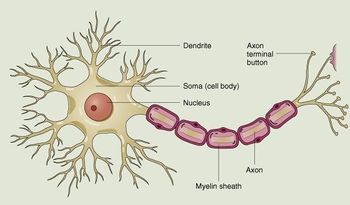
The nerves are the body's messengers. The send the brain's messages and commands to the corresponding parts of the body. The spinal cord is the long bundle of nerves running through the back of your body. It connects with the brain, and all the other nerves. The spinal cord is supported by your backbone. The nerves are made of cells called neurons bound together in bundles called fascicles, nerves, peduncles, or tracts and surrounded by the endoneurium, perineurium, and epineurium membranes. Neurons connect with each other to communicate. Electrochemical waves travel along the axon of one neuron, then move across a gap called a synapse. Sodium and potassium ions are pumped across the synapse to the dendrite of another neuron in a nerve impulse. The image that you see below is a multi polar neuron.
Sense Organs
Sense Organs consist of the eye, ear, nose, tongue
Eye
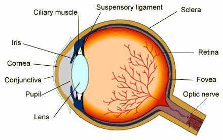
Eye- the organ of vision. It consists of a transparent lens that focuses light on the retina. The retina is covered with light-sensitive cells-rods and cones. The cone cells are sensitive to color and are located in the part of the retina called the fovea, where the light is focused by the lens. The rod cells are not sensitive to color, but have greater sensitivity to light than the cone cells. These cells are located around the fovea and are responsible for peripheral vision and night vision. The eye is connected to the brain through the optic nerve. The optic nerve is called the "blind spot" because it is insensitive to light. The occipital lobe of the brain maps the visual input from the eyes. The brain combines the input of our two eyes into a three-dimensional image. In addition, even though the image on the retina is upside-down because of the focusing action of the lens, the brain compensates and provides the right-side-up perception. In the dark, a substance produced by the rod cells increases the sensitivity of the eye so that it is possible to detect very dim light. In strong light, the iris contracts reducing the size of the aperture that admits light into the eye and a protective obscure substance reduces the exposure of the light-sensitive cells. The spectrum of light to which the eye is sensitive varies from the red to the violet. Lower electromagnetic frequencies in the infrared are sensed as heat, but cannot be seen. Higher frequencies in the ultraviolet and beyond cannot be seen either, but can be sensed as tingling of the skin or eyes depending on the frequency. The human eye is not sensitive to the polarization of light, i.e., light that oscillates on a specific plane.
Ear
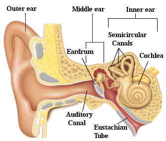
Ear- the organ of hearing. The outer ear protrudes away from the head and is shaped like a cup to direct sounds toward the tympanic membrane, which transmits vibrations to the inner ear through a series of small bones in the middle ear called the malleus, incus and stapes. The inner ear, or cochlea, is a spiral-shaped chamber covered internally by nerve fibers that react to the vibrations and transmit impulses to the brain via the auditory nerve. The brain combines the input of our two ears to determine the direction and distance of sounds. The inner ear has a vestibular system formed by three semicircular canals that are approximately at right angles to each other and which are responsible for the sense of balance and spatial orientation. The inner ear has chambers filled with a viscous fluid and small particles (otoliths) containing calcium carbonate. The movement of these particles over small hair cells in the inner ear sends signals to the brain that are interpreted as motion and acceleration. The human ear can perceive frequencies from 16 cycles per second, which is a very deep bass, to 28,000 cycles per second, which is a very high pitch. The human ear can detect pitch changes as small as 3 hundredths of one percent of the original frequency in some frequency ranges. Some people have "perfect pitch", which is the ability to map a tone precisely on the musical scale without reference to an external standard.
Nose
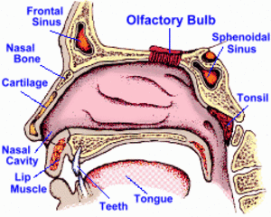
Nose- the organ of smell. The nose is the organ responsible for the sense of smell. The cavity of the nose is lined with mucous membranes that have smell receptors connected to the olfactory nerve. The smells themselves consist of vapors of various substances. The smell receptors interact with the molecules of these vapors and transmit the sensations to the brain. The nose also has a structure called the vomeronasal organ whose function has not been determined, but which is suspected of being sensitive to pheromones that influence the reproductive cycle. The smell receptors are sensitive to seven types of sensations that can be characterized as camphor, musk, flower, mint, ether, acrid, or putrid. The sense of smell is sometimes temporarily lost when a person has a cold.
Tongue
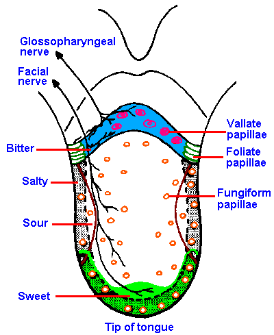
Tongue- the organ of taste. The receptors for taste, called taste buds, are situated chiefly in the tongue, but they are also located in the roof of the mouth and near the pharynx. They are able to detect four basic tastes: salty, sweet, bitter, and sour. The tongue also can detect a sensation called "umami" from taste receptors sensitive to amino acids. Generally, the taste buds close to the tip of the tongue are sensitive to sweet tastes, whereas those in the back of the tongue are sensitive to bitter tastes. The taste buds on top and on the side of the tongue are sensitive to salty and sour tastes. At the base of each taste bud there is a nerve that sends the sensations to the brain. The sense of taste functions in coordination with the sense of smell. The number of taste buds varies substantially from individual to individual, but greater numbers increase sensitivity. The gouge is covered with papillae which makes the tongue have a rough texture.
Spinal Cord
Along with the brain, the spinal cord makes up the central nervous system. It is divided into 31 pairs of nerves, making 62 nerves composed of sensory and motor neurons. The nerves are named off of where they leave the spine. They are divided into 5 groups, cranial, thoracic, lumbar, sacral, and coccygeal.
- 8 pairs in the cervical region
- 12 pairs in the thoracic region
- 5 pairs in the lumbar
- 5 pairs in the sacral
- 1 pair in the coccygeal.
The spinal cord itself in an adult usually ends around the L1 or L2 vertebra, while the remaining axons, forming the cauda equina, continue heading down through the spine and exit where they are supposed to. This happens because the spinal cord stops growing at 4 years of age, while the spine continues to grow. The spinal cord contains both white and gray matter. The white matter travels in tracts to and from the brain. The gray matter forms a sort of h, with 3 horns on either side of the spinal cord. The ventral horn contains somatic motor nuclei, the lateral horn contains autonomic motor nuclei. The dorsal horn is divided, with the farthest back being the somatic sensory portion and the other portion being the visceral sensory portion.
Neural Impulses
Ionic basis of the cellular membrane potential
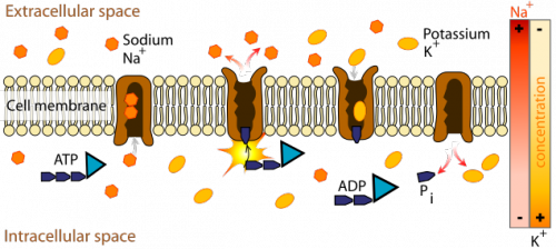
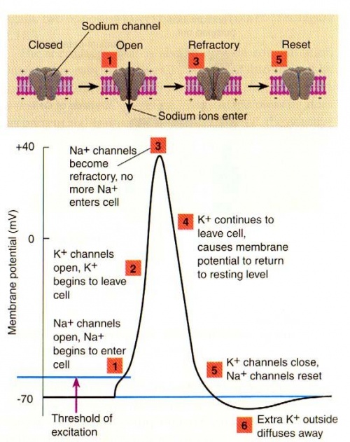
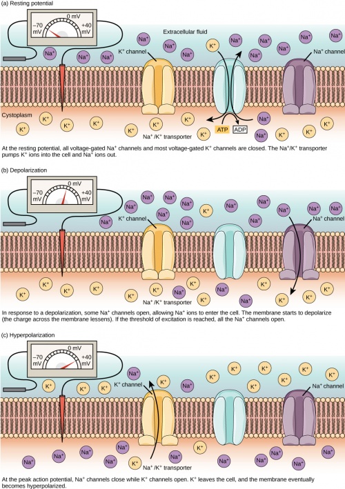
When a neuron is not sending a signal, the inside of the neuron is negative relative to the outside. Although the concentrations of the different ions attempt to balance out on both sides of the membrane, they cannot because the cell membrane allows only some ions to pass through channels (ion channels). At rest, potassium ions (K+) can cross through the membrane easily. Also at rest, chloride ions (Cl-) and sodium ions (Na+) have a more difficult time crossing. The negatively charged protein molecules (A-) inside the neuron cannot cross the membrane. The sodium- potassium pump removes 3 sodium ions for every 2 potassium ions it lets in. When all forces balance out, and the difference in the voltage between the inside and outside of the neuron is measured. That is the resting potential. The resting membrane potential of a neuron is about -70 mV. At rest, there are more sodium ions outside the neuron and more potassium ions inside that neuron.
Action Potential: Generation and Propagation
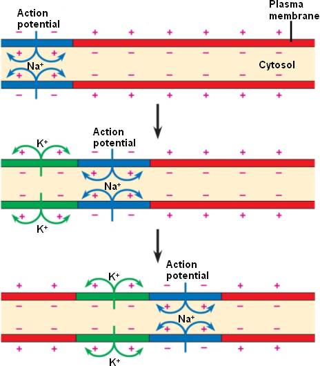
An action potential (spike/ impulse) occurs when a neuron sends information down an axon, away from the cell body.
1. Generation- A stimulus causes the resting potential to move toward 0 mV. When the depolarization reaches about -55 mV a neuron will fire an action potential. If the neuron does not reach this critical threshold level, then no action potential will fire. "ALL OR NONE" principle- The neuron either does not reach the threshold or a full action potential is fired. All sizes of action potentials are the same. Action potentials are caused when different ions cross the neuron membrane. A stimulus first causes sodium channels to open. Because of the concentration gradient, and the inside of the neuron is negative relative to the outside, sodium ions rush into the neuron. Sodium has a positive charge, so the neuron becomes more positive and becomes depolarized. It takes longer for potassium channels to open. When they do open, potassium rushes out of the cell, reversing the depolarization. Sodium channels start to close. This causes the action potential to go back toward -70 mV (a repolarization). The action potential actually goes past -70 mV (a hyperpolarization) because the potassium channels stay open a bit too long. Gradually, the ion concentrations go back to resting levels and the cell returns to -70 mV.
2. Propagation- action potentials are carried from the axon of one neuron to the dendrite of another neuron by synapses and neurotransmitters. Positively charged ions in the fluid outside of the cell flood into the cell and the charge on the inside changes from negative to positive. The electrical impulse moves down the axon.
Impulses From Axon to Dendrite
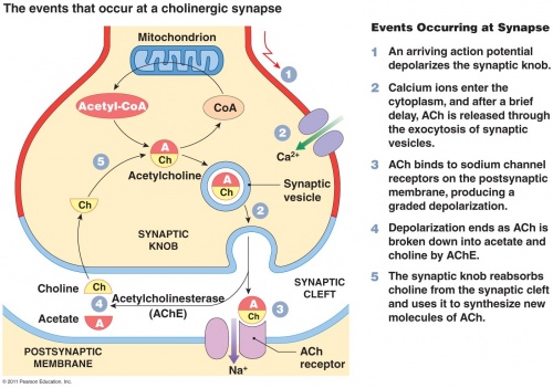
Synapses- junctions where neurons pass signals to other neurons, muscle cells, or gland cells. When a charge reaches a synapse, it may trigger release of tiny bursts of chemicals called neurotransmitters. The synapse contains a small gap separating neurons. The synapse consists of:
- a presynaptic ending that contains neurotransmitters, mitochondria and other cell organelles
- a postsynaptic ending that contains receptor sites for neurotransmitters
- a synaptic cleft or space between the presynaptic and postsynaptic endings.
Neurotransmitters- used to restart impulse in the dendrite of the second neuron. They are chemicals in junctions.
For communication between neurons to occur, an electrical impulse must travel down an axon to the synaptic terminal. At the synaptic terminal, an electrical impulse will trigger the migration of vesicles containing neurotransmitters toward the presynaptic membrane. The vesicle membrane will fuse with the presynaptic membrane releasing the neurotransmitters into the synaptic cleft. The neurotransmitter molecules diffuse across the synaptic cleft where they can bind with receptor sites on the postsynaptic ending to influence the electrical response in the postsynaptic neuron. When a neurotransmitter binds to a receptor on the postsynaptic side of the synapse, it changes the postsynaptic cell's excitability: it makes the postsynaptic cell either more or less likely to fire an action potential. If the number of excitatory postsynaptic events is large enough, they will add to cause an action potential in the postsynaptic cell and a continuation of the "message." Excess neurotransmitters are broken down by enzymes. If they are not, paralysis will occur. One example of a neurotransmitter is acetylcholine. The enzyme that breaks it down is acetylcholinesterase.
Neural Examinations
Encephalography
A neural examination/radiography in which a small amount of cerebrospinal fluid is replaced with a gas.
Magnetic Resonance Imaging (MRI)
A scan of the brain which uses magnets to show images of the body with sharp contrast of soft tissue, air, and bone. This has advantages because it doesn't use radiation.
Disorders/Diseases
Epilepsy
Epilepsy can be described as a "seizure disorder," or a disorder that causes a person to have unprovoked or unexpected seizures. These seizures can seem to have absolutely no cause whatsoever, and after two of these "unprovoked seizures" a person is considered to have epilepsy. Epilepsy can have a varying amount of mental and physical incapacitations, all depending on the person and the severity of the seizures caused by it. Epilepsy Foundation
Alzheimer's Disease
A progressive, degenerative disorder that attacks the neurons, resulting in loss of memory, thinking and language skills, and changes in behavior. The most common form of dementia, it affects memory and comprehension. Most often occurs in people over 65.
Multiple Sclerosis (MS)
A chronic, often disabling disease that attacks the Central Nervous System (CNS). Myelin (fatty substances that surrounds and protects the nerve fibers) is damaged, as well as the nerve fibers themselves. The damaged myelin forms scar tissue (sclerosis), which gives the disease its name. When any part of the myelin sheath or nerve fiber is damaged or destroyed, nerve impulses traveling to and from the brain and spinal cord are distorted or interrupted, producing the variety of symptoms that can occur. Symptoms may be mild, such as numbness in the limbs, or severe, such as paralysis or loss of vision.Considered as an autoimmune disease,
Parkinson's Disease
A chronic and progressive movement disorder which involves malfunction and death of neurons. Some of the neurons create dopamine (chemical that sends messages to brain that controls movement and coordination). As Parkinson's Disease progresses, the neurons die, therefore the amount of dopamine decreases. Primary motor signs include: tremors, bradykinesia (slowness of movement), rigidity, and postural instability (impaired balance and coordination)
Shingles (Herpes Zoster)
Caused by varicella (same virus that causes chickenpox), it mostly occurs in a band or strip on one side of the body called a dermatome, the area where one of the nerves from your spinal cord connects with the skin.
Cerebral Palsy
A disorder that affects muscle tone, movement, and motor skills; usually caused by prenatal brain damage or during first 3-5 years of a child’s life. The brain damage may lead to other health issues including vision, hearing, speech problems, and learning disabilities.
There are three types:
- spastic cerebral palsy – stiffness and movement difficulties,
- athetoid cerebral palsy – involuntary and uncontrolled movements,
- ataxic crebral palsy – disturbed sense of balance and depth perception
Glaucoma
Glaucoma is a group of diseases that can damage the eye's optic nerve and result in vision loss and blindness. While glaucoma can strike anyone, the risk is much greater for people over 60. The most common type of glaucoma is called open-angle glaucoma. In the normal eye, the clear fluid leaves the anterior chamber at the open angle where the cornea and iris meet. When fluid reaches the angle, it flows through a spongy meshwork, like a drain, and leaves the eye.
Sometimes, when the fluid reaches the angle, it passes too slowly through the meshwork drain, causing the pressure inside the eye to build. If the pressure damages the optic nerve, open-angle glaucoma -- and vision loss -- may result.
Pink Eye (Conjunctivitis)
Pink eye (conjunctivitis) is an inflammation or infection of the transparent membrane (conjunctiva) that lines your eyelid and covers the white part of your eyeball. When small blood vessels in the conjunctiva become inflamed, they're more visible. This is what causes the whites of your eyes to appear reddish or pink.
Pink eye is commonly caused by a bacterial or viral infection, an allergic reaction, or — in babies — an incompletely opened tear duct.
Effects of Drugs
Alcohol
Alcohol is a central nervous system depressant. It acts at many sites, including the reticular formation, spinal cord, cerebellum and cerebral cortex, and on many neurotransmitter systems. Alcohol is a very small molecule and is soluble in "lipid" and water solutions. Because of these properties, alcohol gets into the bloodstream very easily and also crosses the blood brain barrier.
In low doses, alcohol:
- produces a relaxing effect
- reduces tension
- lowers inhibitions
- impairs concentration
- slows reflexes
- impairs reaction time
- reduces coordination
In medium doses, alcohol:
- slurs speech
- causes drowsiness
- alters emotions
In high doses, alcohol:
- induces vomiting
- creates breathing difficulties
- unconsciousness
- comas
Caffeine
Caffeine is a alkaloid which acts as a stimulant drug. In humans, it temporarily stimulates the central nervous system, increasing alertness and attention. However, it can cause addiction. If one takes in too much caffeine, they can develop a buildup, called tolerance, in which the caffeine's effects are reduced, and therefore more is needed to create the same effects as when there was less tolerance.
Nicotine
Nicotine (also spelled nicotene) is a alkaloid which constitutes about 0.6-3% of tobacco plants. As well as being a chemical, which, when contained in plants, resists animal consumption, it is also a stimulant for mammals. About 1 milligram is contained in the average cigarette, and it is responsible for the addiction to smoking. When nicotine enters the body, it quickly enters the bloodstream. When it binds to receptors in the central nervous system, they "turn up the volume" on neurotransmitters.
Marijuana
Marijuana comes from a plant called "cannabis sativa"; the chemical in the plant that produces the altered states of consciousness is called "delta-9 tetrahydrocannabinol" or "THC". THC acts on cannabinoid receptors, which are found on neurons in many places in the brain. These areas are involved in memory (hippocampus), concentration (cerebral cortex), perception (sensory portions of the cerebral cortex), and movement (cerebellum, substantia nigra, globus pallidus).
In low-medium doses, marijuana:
- causes relaxation
- reduces coordination
- reduces blood pressure
- induces sleepiness
- disrupts attention
- creates an altered sense of time and space
In high doses, marijuana:
- induces hallucinations
- creates delusions
- impairs memory
- provokes disorientation
Event Tips
- Abbreviate where possible on your resource sheet-saves space!
- Check out the official Science Olympiad website for general resources.


