Anatomy/Muscle List
Jump to navigation
Jump to search
This is a list of muscles tested on in the Muscular System portion of Anatomy and Physiology. See the official 2016 list here: [1].
Muscles of the Head and Neck
| Muscle Name | Origin | Insertion | Function | Picture |
|---|---|---|---|---|
| Frontalis | Occipital : highest nuchal line and mastoid process. Frontal: superior fibers of upper facial muscles | Galeal aponeurosis (skin above the eyebrows) | Wrinkles forehead and fixes galeal aponeurosis | |
| Orbicularis oris | Near midline on anterior surface of maxilla and mandible and modiolus at angle of mouth | Mucous membrane of margin of lips and raphe with buccinator at modiolus; skin around lips | Narrows orifice of mouth, purses lips and puckers lip edges | |
| Orbicularis oculi | Medial orbital margin and lacrimal sac (orbital, palpebral and lacrimal parts) | Lateral palpebral raphe | Closes eyelids, aids passage and drainage of tears | |
| Occipitofrontalis | 2 occipital bellies and 2 frontal bellies; occipital bone | Galea aponeurotica | Raises eyebrows, wrinkles forehead | |
| Zygomaticus major | Anterior surface of zygomatic bone | Modiolus at angle of mouth | Elevates and draws angle of mouth laterally | |
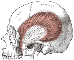
| Masseter | Anterior two thirds of zygomatic arch and zygomatic process of maxilla | Lateral surface of angle and lower ramus of mandible | Elevates mandible (enables forced closure of mouth) | |
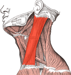
| Sternocleidomastoid | Anterior and superior manubrium and superior medial third of clavicle | Lateral aspect of mastoid process and anterior half of superior nuchal line | Flexes and laterally rotates cervical spine. Protracts head when acting together. Extends neck when neck already partially extended | |
| Trapezius | Medial third superior nuchal line, ligament nuchae, spinous processes and supraspinous ligaments to T12 | Upper fibers to lateral third of posterior border of clavicle; lower to medial acromion and superior lip of spine of scapula to deltoid tubercle | Laterally rotates, elevates and retracts scapula. If scapula is fixed, extends and laterally flexes neck | |
| Buccinator | External alveolar margins of maxilla and mandible by molar teeth, to maxillary tubercle and pterygoid hamulus and posterior mylohyoid line respectively, then via pterygomandibular raphe between bones | Decussates at modiolus of mouth and interdigitates with opposite side | Aids mastication, tenses cheeks in blowing and whistling, aids closure of mouth |
Muscles That Move the Upper Extremities
| Muscle Name | Origin | Insertion | Function | Picture |
|---|---|---|---|---|
| Pectoralis major | Clavicular head Clavicular head-medial half clavicle. Sternocostal head-lateral manubrium and sternum, six upper costal cartilages and external oblique aponeurosis | Lateral lip of bicipital groove of humerus and anterior lip of deltoid tuberosity | Clavicular head:flexes and adducts arm. Sternal head: adducts and medially rotates arm. Accessory for inspiration | |
| Latissimus dorsi | Spine T7, spinous processes and supraspinous ligaments of all lower thoracic, lumbar and sacral vertebrae, lumbar fascia, posterior third iliac crest, last four ribs (interdigitating with external oblique abdominis) and inferior angle of scapula | Floor of bicipital groove of humerus after spiraling around teres major | Extends, adducts and medially rotates arm. Costal attachment helps with deep inspiration and forced expiration | |
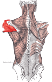
| Deltoid | Lateral third of clavicle, acromion, spine of scapula to deltoid tubercle | Middle of lateral surface of humerus (deltoid tuberosity) | Abducts arm, anterior fibers flex and medial rotate, posterior fibers extend and lateral rotate | |
| Teres major | Oval area (lower third) of lateral side of inferior angle of scapula below teres minor | Medial lip of bicipital groove of humerus | Medially rotates and adducts arm. Stabilizes shoulder joint | |
| Biceps brachii | Long head:supraglenoid tubercle of scapula. Short head: coracoid process of scapula with coracobrachialis | posterior border of bicipital tuberosity of radius (over bursa) and bicipital aponeurosis to deep fascia and subcutaneous ulna | flexes elbow and supinates forearm | |
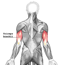
| Triceps brachii | Long head: infraglenoid tubercle of scapula. lateral head: upper half posterior humerus (linear origin). medial head: lies deep on lower half posterior humerus inferomedial to spiral groove and both intermuscular septa | Posterior part of upper surface of olecranon process of ulna and posterior capsule | Extends elbow. Long head stabilizes shoulder joint. medial head retracts capsule of elbow joint on extension | |
| Brachialis | Anterior lower half of humerus and medial and lateral intermuscular septa | Coronoid process and tuberosity of ulna | Flexes elbow | |
| Brachioradialis | Upper two thirds of lateral supracondylar ridge of humerus and lateral intermuscular septum | Base of styloid process of radius | Flexes arm at elbow and brings forearm into midprone position | |
| Palmaris longus | Common flexor origin of medial epicondyle of humerus | Flexor retinaculum and palmar aponeurosis | Flexes wrist and tenses palmar aponeurosis | |
| Flexor carpi radialis | Common flexor origin of medial epicondyle of humerus | Bases of 2nd and 3rd metacarpals via groove in trapezium and slip to scaphoid | Flexes and abducts wrist | |
| Flexor digitorum superficialis | Humeral head: common flexor origin of medial epicondyle humerus, medial ligament of elbow. Ulnar head: medial border of coronoid process and fibrous arch. Radial head: whole length of anterior oblique line | Tendons split to insert onto sides of middle phalanges of medial four fingers | Flexes proximal interphalangeal joints and secondarily metacarpophalangeal joints and wrist | |
| Extensor carpi radialis brevis | Common extensor origin on anterior aspect of lateral epicondyle of humerus | Posterior base of 3rd metacarpal | Extends and abducts hand at wrist | |
| Extensor carpi radialis longus | Lower third of lateral supracondylar ridge of humerus and lateral intermuscular septum | Posterior base of 2nd metacarpal | Extends and abducts hand at wrist | |
| Extensor digitorum | Common extensor origin on anterior aspect of lateral epicondyle of humerus | External expansion to middle and distal phalanges by four tendons. Tendons 3 and 4 usually fuse and little finger just receives a slip | Extends all joints of fingers | |
| Extensor digiti minimi | Common extensor origin on anterior aspect of lateral epicondyle of humerus | Extensor expansion of little finger-usually two tendons which are joined by a slip from extensor digitorum at metacarpophalangeal joint | Extends all joints of little finger | |
| Extensor carpi ulnaris | Common extensor origin on anterior aspect of lateral epicondyle of humerus | Base of 5th metacarpal via groove by ulnar styloid | Extends and adducts hand at wrist |
Extra Images
Muscles of the Trunk
| Muscle Name | Origin | Insertion | Function | Picture |
|---|---|---|---|---|
| External Intercostals | Inferior border of ribs as far back as posterior angles | Superior border of ribs below, passing obliquely downwards and backwards | Fix intercostal spaces during respiration. Aids forced inspiration by elevating ribs | |
| Internal Intercostals | Inferior border of ribs as far back as posterior angles | Superior border of ribs below , passing obliquely downwards and backwards | Fix intercostal spaces during respiration. Aids forced inspiration by elevating ribs | |
| Innermost Intercostals (not techically on the list, but good to know anyway) | Internal aspect of ribs above and below | Internal aspect of ribs above and below | Fix intercostal spaces during respiration | |
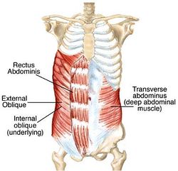
| Transverse abdominis | Costal margin, lumbar fascia, anterior two thirds of iliac crest and lateral half of inguinal ligament | Aponeurosis of posterior and anterior rectus sheath and conjoint tendon to pubic crest and pectineal line | Supports abdominal wall, aids forced expiration and raising intra-abdominal pressure. Conjoint tendon supports posterior wall of inguinal canal | |
| Infraspinatus | Medial three quarters of infraspinous fossa of scapula and fibrous intermuscular septa | Middle facet of greater tuberosity of humerus and capsule of shoulder joint | Laterally rotates arm and stabilizes shoulder joint | |
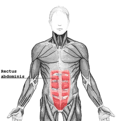
| Rectus abdominis | Pubic crest and pubic symphysis | 5, 6, 7 costal cartilages, medial inferiorcostal margin and posterior aspect of xiphoid | Flexes trunk, aids forced expiration and raise intra-abdominal pressure | |
| Serratus anterior | Upper eight ribs and anterior intercostal membranes from midclavicular line. Lower four interdigitating with external oblique | Inner medial border scapula. 1 and 2: upper angle; 3 and 4: length of costal surface ; 5-8: inferior angle | Laterally rotates and protracts scapula | |
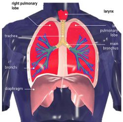
| Diaphragm | Vertebral:crura from bodies of L1, 2 (left), L1-3 (right). Costal: medial and lateral arcuate ligs, inner aspect of lower six ribs. Sternal: two slips from posterior aspect of xiphoid | Central tendon | Inspiration and assists in raising intra-abdominal pressure |
Muscles That Move the Lower Extremities
| Muscle Name | Origin | Insertion | Function | Picture |
|---|---|---|---|---|
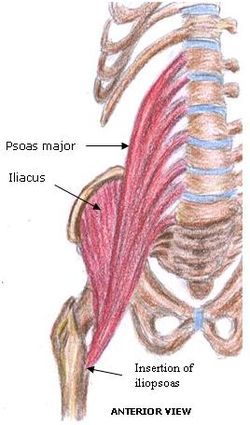
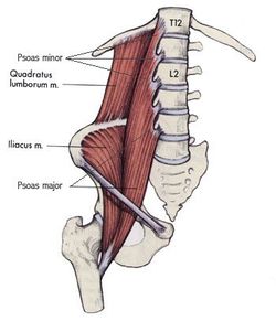
| Iliopsoas(made up of 3 muscles: see below) | ||||
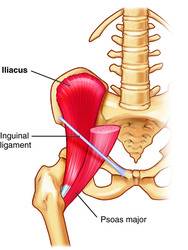
| Iliacus | Iliac fossa within abdomen | Lowermost surface of lesser trochanter of femur | Flexes medially rotates hip | |
| Psoas major | Transverse processes of L1-5, bodies of T12-L5 and intervertebral discs below bodies of T12-L4 | Middle surface of lesser trochanter of femur | Flexes and laterally rotates hip | |
| Psoas minor | Bodies of T12 and L1 and intervening intervertebral disc | Fascia over psoas major and iliacus | Weak flexor of trunk) | |
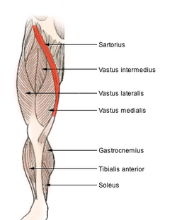
| Sartorius | Immediately below anterior superior iliac spine | Upper medial surface of shaft of tibia | Flexes, abducts, laterally rotates thigh at hip. Flexes, medially rotates leg at knee | |
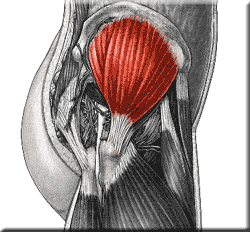
| Gluteus maximus | Outer surface of ilium behind posterior gluteal line and posterior third of iliac crest lumbar fascia, lateral mass of sacrum, sacrotuberous ligament and coccyx | Deepest quarter into gluteal tuberosity of femur, remaining three quarters into iliotibial tract (anterior surface of lateral condyle of tibia) | Extends and laterally rotates hip. Maintains knee extended via iliotibial tract; chief antigravity muscle when sitting | |
| Gluteus medius | Outer surface of ilium between posterior and middle gluteal lines | Posterolateral surface of greater trocanter of femur | Abducts and medially rotates hip. Tilts pelvis when walking | |
| Tensor fasciae latae | Outer surface of anterior iliac crest between tubercle of the iliac crest and anterior superior iliac spine | Iliotibial tract (anterior surface of lateral condyle of tibia) | Maintains knee extended (assists gluteus maximus) and abducts hip | |
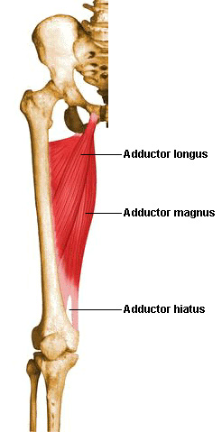
| Adductor longus | Body of pubis inferior and medial to pubic tubercle | Lower two thirds of medial linea aspera | Adducts and medially rotates hip | |
| Gracilis | Outer surface of ischiopubic ramus | Upper medial shaft of tibia below sartorius | Adducts hip. Flexes knee and medially rotates flexed knee | |
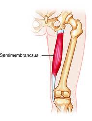
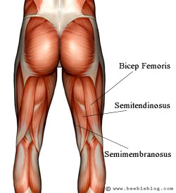
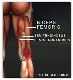
| Semimembranosus | Upper outer quadrant of posterior surface of ischial tuberosity | Medial condyle of tibia below articular margin, fascia over popliteus and oblique popliteal ligament | Flexes and medially rotates knee. Extends hip | |
| Semitendinosus | Upper inner quadrant of posterior surface of ischial tuberosity | Upper medial shaft of tibia below gracilis | Flexes and medially rotates knee. Extends hip | |
| Biceps femoris | Long head: upper inner quadrant of posterior surface of ischial tuberosity. Short head:middle third of linea aspera, lateral supracondylar ridge of femur | Styloid process of head of fibula. lateral collateral ligament and lateral tibial condyle | Flexes and laterally rotates knee. Long head extends hip | |
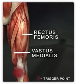
| Rectus femoris | Straight head: anterior inferior iliac spine. Reflected head: ilium above acetabulum | Quadriceps tendon to patella , via ligamentum patellae into tubercle of tibia | Extends leg at knee. Flexes thigh at hip | |
| Vastus lateralis | Upper intertrochanteric line, base of greater trochanter, lateral linea aspera, lateral supracondylar ridge and lateral intermuscular septum | Lateral quadriceps tendon to patella, via ligamentum patellae into tubercle of tibia | Extends knee | |
| Vastus intermedius | Anterior and lateral shaft of femur | Quadriceps tendon to patella, via ligamentum patellae into tubercle of tibia | Extends knee | |
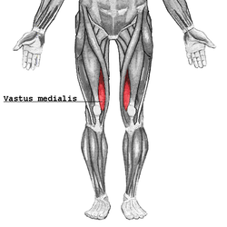
| Vastus medialis | Lower intertrochanteric line, spiral line, medial linea aspera and medial intermuscular septum | Medial quadriceps tendon to patella and directly into medial patella, via ligamentum patellae into tubercle of tibia | Extends knee. Stabilizes patella | |
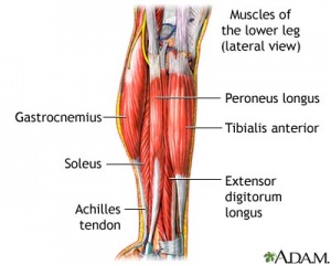
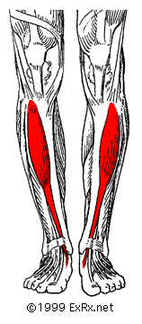
| Tibialis anterior | Upper half of lateral shaft of tibia and interosseous membrane | Inferomedial aspect of medial cuneiform and base of 1st metatarsal | Extends and inverts foot at ankle. Holds up medial longitudinal arch of foot | |
| Gastrocnemius | Lateral head: posterior surface of lateral condyle of femur and highest of three facets on lateral condyle. medial head: posterior surface of femur above medial condyle | Tendo calcaneus to middle of three facets on posterior aspect of calcaneus | Plantar flexes foot. Flexes knee | |
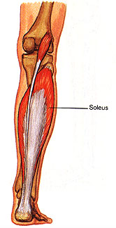
| Soleus | Soleal line and middle third of posterior border of tibia and upper quarter of posterior shaft of fibula including neck | Tendo calcaneus to middle of three facets on posterior surface of calcaneus | Plantar flexes foot (aids venous return) | |
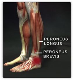
| Peroneus longus | Upper two thirds of lateral shaft of fibula, head of fibula and superior tibiofibular joint | Plantar aspect of base of 1st metatarsal and medial cuneiform, passing deep to long plantar ligament | Plantar flexes and everts foot. Supports lateral longitudinal and transverse arches | |
| Peroneus brevis | Lower two thirds lateral shaft of fibula | Tuberosity of base of 5th metatarsal | Plantar flexes and everts foot. Supports lateral longitudinal arch |
Previously Listed Muscles
ExpandPreviously Listed Muscles (not on the 2016 list)
|


